A medical student-run newsletter showcasing the latest cutting edge research in otolaryngology
Issue #102
17 December 2025
Educational Pearl
Epiglottitis
Overview: Epiglottitis is inflammation of the epiglottis and surrounding supraglottic structures, most often due to infection, that can rapidly progress to life-threatening upper airway obstruction without timely intervention. Airway protection is critical, especially in children, whose distress and agitation can quickly worsen obstruction.
Epidemiology:
Adults: ~0.6 - 1.9 cases per 100,000 per year, usually in those > 45 years old
Common comorbidities include hypertension, diabetes, end-stage kidney disease, substance use disorder, and immunodeficiency
Increased disease severity has been associated with BMI > 25, diabetes, concurrent pneumonia, epiglottic cysts
Children: ~2 cases per 10,000,000 per year in areas with high vaccination rates
Widespread Haemophilus influenzae type b vaccination decreased incidence in kids
Etiology:
Most often due to infection
Rich lymphatic and vascular supply of epiglottis facilitates rapid infection spread
Haemophilus influenzae type b = most common infectious cause
Non-Infectious Causes:
Physical trauma, thermal injury (inhalation burns from e-cigarettes, hot liquids), caustic ingestion, granulomatous disease, angioedema
Diagnosis:
Clinical Presentation:
Acute onset (within hours)
Upper airway narrowing or obstruction secondary to supraglottic edema can present with stridor, drooling, “hot potato” voice, severe sore throat, dysphagia
Evaluation:
Clinical suspicion based on the above presentation is key
Endoscopic examination shows inflamed supraglottis +/- false vocal cord edema
Lateral neck x-ray may assist when suspicion is intermediate
Look for classic “thumb sign” (enlarged, swollen epiglottis)
Laboratory Studies:
CBC, blood culture, epiglottic culture if intubated
Avoid if airway obstruction is imminent
Management:
Goal: Secure the airway
Total or Near-Total Airway Obstruction:
Bag-mask ventilation with 100% FiO2
Rapid sequence intubation with surgeon available for surgical airway if needed
Directly lift epiglottis during laryngoscopy if visualization is poor
Medical Therapy:
Start empiric intravenous antibiotics covering Haemophilus influenzae type b, Streptococcus pyogenes, Streptococcus pneumoniae, Staphylococcus aureus
Glucocorticoids/racemic epinephrine have no proven benefit (not recommended)
Further Readings:
[1] StatPearls - Epiglottitis
[2] Acute Epiglottitis Trends, Diagnosis, Management
Source:
[1] UpToDate - Epiglottitis Clinical Features + Diagnosis
Educational Pearl written by Gina Spencer
Queen's University School of Medicine
Question of the Week
A 47-year-old man comes to the clinic for evaluation of unusual facial symptoms that occur during meals. Eight months ago, he underwent a left superficial parotidectomy for a pleomorphic adenoma. His postoperative course was uncomplicated, and he has had no issues with facial movement or sensation. Over the past several months, the patient reports that within seconds of tasting or smelling food, he develops a warm sensation followed by flushing and sweating over the area just anterior to his left ear. The symptoms last for the meal duration and resolve shortly afterward. He denies fevers, facial pain, weakness, numbness, xerostomia, or dysphagia. On examination, facial nerve function is intact and the left preauricular surgical scar is well healed. Biting into a lemon wedge produces localized erythema and moisture along the incision line.
Which of the following best explains this patient’s symptoms?
(A) Aberrant sympathetic fiber regeneration to sweat glands
(B) Aberrant parasympathetic secretomotor fiber regeneration from auriculotemporal nerve to sweat glands
(C) Regeneration of facial nerve branches to sensory fibers
(D) Loss of sympathetic tone with unopposed parasympathetic activity
(E) Cross-innervation of glossopharyngeal fibers to buccal glands
Question of the Week written by Adriana Báez Berríos
Icahn School of Medicine at Mount Sinai
· · · · · · · · · · · · · · · · · · · · · · · · · · · · · · · · · · · · · · · · · · · · · · · · · · · · · · · · · · · · · · · ·
Looking for the answer to this Question of the Week?
Find it at the bottom of this newsletter!
The high-quality otolaryngology content we deliver each month is made possible by the dedication of our national faculty reviewers. We sincerely thank them for their contributions to The Auricle.
Facial Plastic and Reconstructive Surgery
Dr. Jacob Dey, MD
Mayo Clinic - Minnesota
Head and Neck Surgery
Dr. Akina Tamaki, MD
Lewis Katz School of Medicine at Temple University
Laryngology / Med Student Feature
Dr. Inna Husain, MD
Powers Health
Otology and Neurotology
Dr. Mohamed Elrakhawy, MD
Rush University Medical Center
Rhinology and Skull Base Surgery
Dr. Peter Papagiannopoulos, MD
Rush University Medical Center
Sleep Surgery
Dr. Rheena Dhanda Patil, MD
University of Cincinnati Medical Center
Medical Student Feature Series
This series showcases student-led research published in leading otolaryngology journals, highlighting evidence-based discoveries from the next generation of otolaryngologists.

Ganesan V, Zhao R, Li A, Tai K, Rameau A. Association of Cognition, Frailty, & Peak Airflow With Dysphagia in Older Adults: A National Study. Laryngoscope. Published online August 9, 2025. [Article Link]
Is dysphagia hiding in frailty, cognition, or airflow?
Dysphagia, a subjective difficulty with swallowing distinct from clinician-rated swallowing impairment, is common among older adults and associated with adverse health outcomes. This cross-sectional study used Round 12 (2022 to 2023) of the National Health and Aging Trends Study to evaluate 5899 Medicare beneficiaries aged ≥ 65 years, defining dysphagia by self-report or use of rehabilitation services for chewing or swallowing problems. Primary exposures included frailty status (robust, pre-frail, frail), cognitive status (normal cognition, cognitive impairment, dementia), and peak expiratory flow (PEF; normal vs. low), assessed using validated methods, with associations evaluated using multivariable logistic regression adjusted for age, sex, race, ethnicity, and sampling weights. Compared with robust older adults, pre-frail and frail individuals had significantly higher odds of self-reported dysphagia (pre-frail: adjusted odds ratio [aOR] = 3.45, 95% confidence interval [CI] 2.4 to 5.2; frail: aOR = 7.57, 95% CI 5.1 to 11.7; both p ≤ 0.001), and low PEF remained independently associated with dysphagia after adjustment for frailty (aOR = 1.73, 95% CI 1.4 to 2.2, p ≤ 0.001; aOR = 1.46, 95% CI 1.2 to 1.8). Dementia was not associated with self-reported dysphagia compared with normal cognition (aOR = 1.35; p = 0.13), whereas low PEF was strongly associated with frailty (frail vs. robust: aOR = 5.25, 95% CI 3.8 to 7.3; p ≤ 0.001). These findings suggest that frailty status and simple peak flow measurements may help identify older adults at higher risk for dysphagia and aspiration, supporting their integration into routine dysphagia screening and multidisciplinary geriatric assessment. The absence of an association between dementia and self-reported dysphagia highlights the likelihood of under-reporting in cognitively impaired patients and the need for objective swallow assessments rather than reliance on symptoms alone in this population.
Medical Student Feature Summary written by Priyanka Shah
Edward Via College of Osteopathic Medicine
Vinayak Ganesan
Case Western Reserve University School of Medicine
Class of 2029
Cognition, Frailty, and Airflow vs. Dysphagia
Are you a medical student with a recent first-author publication?
Email theauricleotolaryngology@gmail.com to be featured!
Facial Plastic and Reconstructive Surgery
Endoscopic Versus Pretrichial Brow Lift Outcomes
Garg N, Alnemri A, DeKloe J, et al. A Comparison of Brow Elevation and Complications: Endoscopic Versus Pretrichial Brow Lift. Laryngoscope. Published online November 14, 2025. [Article Link]
Two techniques, one long-term outcome
Brow ptosis contributes to significant functional and cosmetic concerns, particularly in the aging population, yet the optimal surgical approach to brow lift remains debated due to limited long-term comparative data. This retrospective study evaluated 72 adult patients undergoing cosmetic brow lift (mean age 63.3 years, range 39 to 78 years; N = 67, 93.1% female; N = 5, 6.9% male), comparing endoscopic (N = 50, 69.4%) and pretrichial (N = 22, 30.6%) techniques and associated postoperative complication profiles. Standardized full-face frontal photographs with relaxed forehead musculature were analyzed using a validated ImageJ brow elevation ratio, calculated as vertical brow height from the intercanthal line to the superior eyebrow border divided by the horizontal distance from the lateral limbus to the medial canthus. The endoscopic approach achieved greater immediate postoperative elevation at one week (+20.8% vs. +12.8%; p < 0.01), while both techniques demonstrated comparable long-term brow height retention beyond six months (mean follow-up 429 ± 278 days, elevation ratio 1.38 vs. 1.23; p > 0.05) and similar interval declines over time (-2.6% vs. -2.9%; p > 0.05). Rates of postoperative complications, including brow weakness, paresthesia, alopecia, and suboptimal scarring, did not differ between groups (all p > 0.05). These findings suggest that brow lift technique selection should be individualized based on hairline position, forehead length, hair density, and aesthetic goals, as long-term durability and safety outcomes appear equivalent between approaches.
Facial Plastic and Reconstructive Surgery Summary written by Ashton Huppert Steed
University of Arizona College of Medicine Phoenix
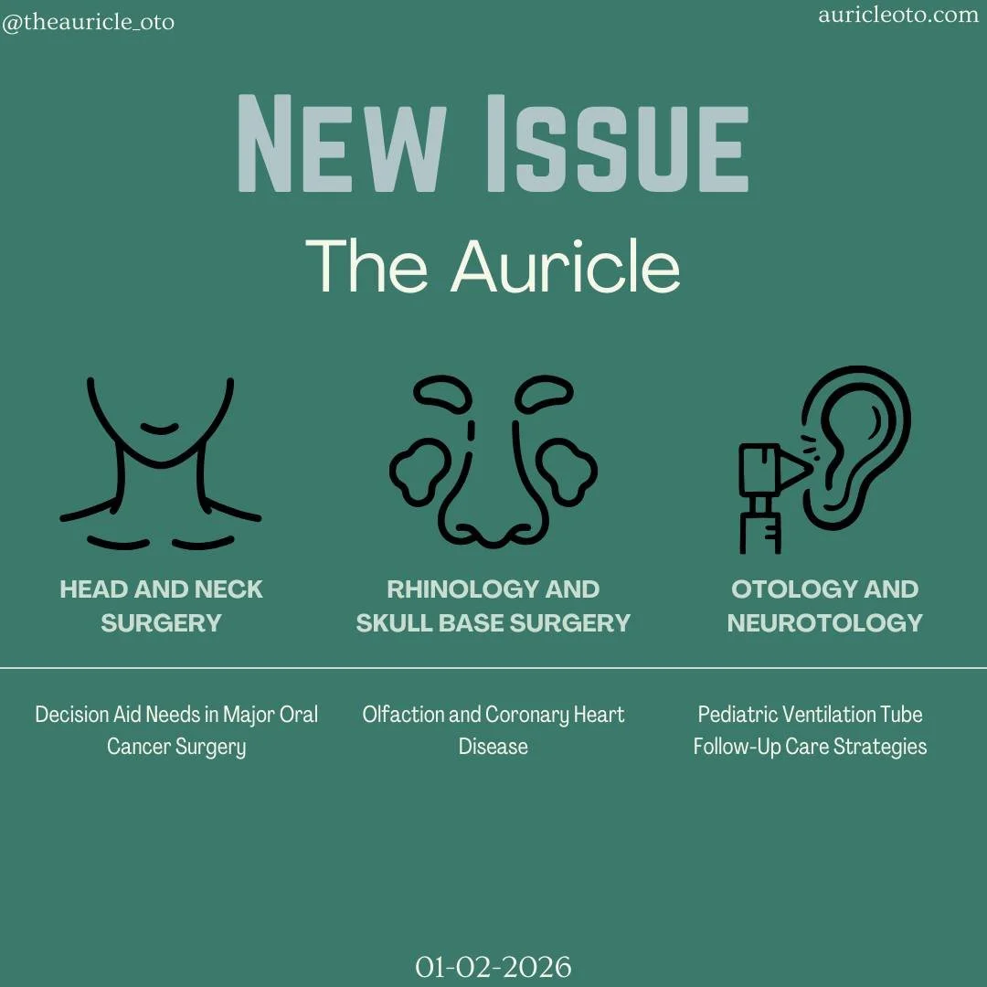
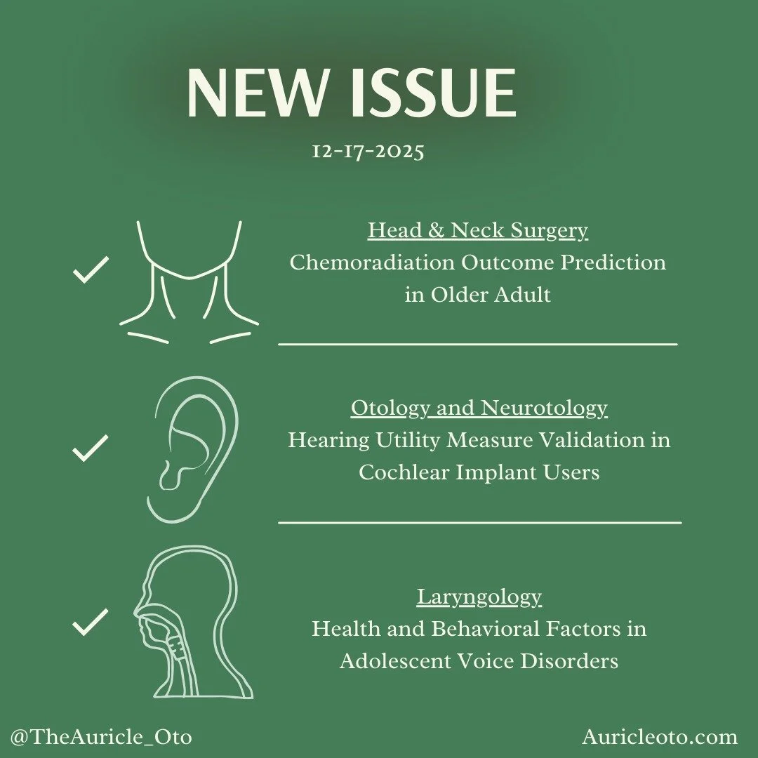
Head and Neck Surgery
Chemoradiation Outcome Prediction in Older Adults
Marschner SN, Lombardo E, Haehl E, et al. Outcome Prediction in Older Adults With Head and Neck Cancer Undergoing Chemoradiation. JAMA Otolaryngol Head Neck Surg. Published online November 6, 2025. [Article Link]
Older adults with head and neck squamous cell carcinoma (HNSCC) are underrepresented in clinical trials, limiting the availability of evidence-based guidance for treatment planning in this population. This international retrospective study used data from the SENIOR registry to develop artificial neural network (ANN) models predicting overall survival (OS) and progression-free survival (PFS) in adults aged ≥ 65 years with locoregionally advanced HNSCC treated with definitive chemoradiation. Separate ANN models were trained for OS (N = 898) and PFS (N = 945), achieving moderate discriminative performance (OS: receiver operating characteristic–area under the curve [ROC-AUC] = 0.68, 95% confidence interval [CI] 0.60 to 0.76; PFS: ROC-AUC = 0.64, 95% CI 0.56 to 0.72). Based on mean Shapley additive explanation (SHAP) values, the strongest predictors of high-risk status in the OS model included negative human papillomavirus (HPV) status (0.048 ± 0.026), decreased estimated glomerular filtration rate (0.046 ± 0.045), poor Eastern Cooperative Oncology Group performance status (0.043 ± 0.040), and advanced nodal classification (0.039 ± 0.027), whereas the PFS model was primarily influenced by negative HPV status (0.107 ± 0.087), higher age-adjusted Charlson Comorbidity Index score (0.091 ± 0.066), elevated C-reactive protein levels (0.084 ± 0.055), and advanced nodal classification (0.076 ± 0.046). These findings suggest that ANN-based models can stratify older adults with HNSCC into prognostic risk groups and support more personalized chemoradiation decision-making in this underrepresented population.
Head and Neck Surgery Summary written by Brooke Swain
Vanderbilt University School of Medicine
Laryngology
Health and Behavioral Factors in Adolescent Voice Disorders
Fujiki RB, Thibeault SL. Medical Comorbidities and Behavioral Health in Adolescents With Voice Disorders. JAMA Otolaryngol Head Neck Surg. 2025;151(10):913-922. [Article Link]
Voice disorders are relatively common in the United States, affecting approximately 6% of adults and 7.4% of adolescents. This cross-sectional study evaluated medical, behavioral health, and medication-related factors associated with dysphonia in adolescents using survey data from 502 participants aged 13 to 17 years (mean age 15.2 ± 1.27 years). Dysphonia was defined by self-report as any voice problem that interfered with communication, with data collected on medical diagnoses, behavioral health conditions, and medication use. Multivariable logistic regression identified several medical comorbidities associated with higher odds of dysphonia, including a family history of voice disorders (odds ratio [OR] = 7.3, 95% confidence interval [CI] 3.2 to 14.7), cancer (OR = 6.5, 95% CI 2.9 to 10.9), diabetes (OR = 3.4, 95% CI 2.1 to 5.1), neurologic disorders (OR = 3.1, 95% CI 1.9 to 5.1), gastrointestinal conditions (OR = 2.5, 95% CI 1.8 to 3.2), and acid reflux (OR = 2.3, 95% CI 1.5 to 3.6). Medication use, including inhalers (OR = 1.6, 95% CI 1.2 to 2.5), antidepressant or anxiolytic medications (OR = 3.1, 95% CI 1.9 to 4.2), and steroids or hormonal therapies (OR = 4.1, 95% CI 1.9 to 8.2), was also significantly associated with dysphonia. From a behavioral health perspective, anxiety (OR = 2.2, 95% CI 1.8 to 3.7) and depression (OR = 1.9, 95% CI 1.5 to 2.9) were independently associated with voice problems, with greater anxiety severity correlating with increased dysphonia risk. These findings suggest that adolescent dysphonia is multifactorial, with contributions from medical comorbidities, behavioral health conditions, and medication exposure, highlighting the need for further research to clarify causal risk factors and inform targeted prevention strategies.
Laryngology Summary written by Maaz Haji
Chicago Medical School, Rosalind Franklin University
Otology and Neurotology
Hearing Utility Measure Validation in Cochlear Implant Users
Dixon PR, Shah AH, McRackan TR, et al. Validation of the Hearing Utility Measure (HUM) in Cochlear Implant Candidates and Users. Otolaryngol Head Neck Surg. 2025; 173(6):1470-1477. [Article Link]
Generic health utility instruments, such as the Health Utilities Index Mark-3 (HUI-3), inadequately capture hearing-specific outcomes, including sound localization, tinnitus, and music appreciation, limited accurate cost-effectiveness analyses for hearing interventions such as cochlear implantation. This prospective cohort study conducted at a tertiary academic center in Ontario, Canada enrolled 1315 adult cochlear implant candidates (median age 64 years, interquartile range 51 to 75 years; N = 687, 52.2% female; N = 628, 47.8% male), excluding patients undergoing revision surgery for device failure or complications, to validate the newly developed Hearing Utility Measure (HUM), which assesses hearing across seven domains. Participants completed the HUM alongside established instruments, including the Cochlear Implant Quality of Life Profile-35 (CIQOL-35), Speech, Spatial and Qualities of Hearing Scale-49 (SSQ-49), Tinnitus Handicap Inventory, and audiometric testing at cochlear implant evaluation and 12 months post-activation. The HUM demonstrated excellent test-retest reliability and strong construct validity (intraclass correlation coefficient = 0.90, 95% confidence interval [CI] 0.83 to 0.94), with robust correlations with CIQOL-35 and SSQ-49 (CIQOL-35: r = 0.69, 95% CI 0.66 to 0.72; SSQ-49: r = 0.70, 95% CI 0.67 to 0.73). Known-groups validity showed significantly higher HUM scores among single-sided deafness (SSD) candidates compared with traditional or asymmetric hearing loss candidates (0.76 vs. 0.58; p = 0.002 and p < 0.001, respectively). Following cochlear implantation, HUM scores improved significantly in both SSD and traditional recipients (SSD: 0.70 to 0.78, p = 0.002; traditional: 0.57 to 0.75; p < 0.001), whereas HUI-3 failed to detect hearing utility improvements in SSD recipients (1.0 to 0.71; p = 0.900). These findings demonstrate that the HUM captures clinical meaningful hearing-specific outcomes overlooked by generic instruments and enables more accurate cost-effectiveness analyses to inform cochlear implant policy and decision-making.
Otology and Neurotology Summary written by Rushi Vekariya
University of Central Florida College of Medicine
Rhinology and Skull Base Surgery
Sex vs. Chronic Rhinosinusitis Diagnosis and Biomarkers
Chiu RG, Ahn A, Eldeirawi K, et al. Biological Sex and Chronic Rhinosinusitis Diagnosis and Biomarkers. JAMA Otolaryngol Head Neck Surg. Published online October 23, 2025. [Article Link]
His & Hers sinuses: Sex-based differences in CRS phenotypes
Biological sex influences immune and inflammatory pathways, yet its role in chronic rhinosinusitis (CRS) phenotypes remains incompletely understood. This cross-sectional study analyzed data from a nationwide United States database including 258,245 participants to evaluate whether sex at birth is independently associated with CRS diagnosis, specifically CRS without nasal polyps (CRSsNP) and CRS with nasal polyps (CRSwNP). Primary outcomes included CRS subtype, with secondary analyses assessing serum eosinophils, immunoglobulin E (IgE), neutrophils, and uric acid, adjusting for demographic, socioeconomic, and clinical covariates. Female participants demonstrated a higher overall prevalence of CRS than male participants (12.1% females vs. 8.6% males), driven by higher rates of CRSsNP (11.4% females vs. 7.6% males), whereas CRSwNP was more prevalent among men (1.0% males vs. 0.7% females). After multivariable adjustment, female sex was associated with increased odds of CRSsNP in individuals younger than 60 years (odds ratio [OR] = 1.44, 95% Holm-Bonferroni-corrected confidence interval [CIH-B] 1.35 to 1.54) and those aged 60 years or older (OR = 1.32, 95% CIH-B 1.23 to 1.40), but reduced odds of CRSwNP (OR = 0.63, 95% CIH-B 0.52 to 0.76). Biomarker analyses showed lower serum eosinophil counts and lower IgE levels among females with CRSsNP (serum eosinophil counts: β = -0.35, 95% CIH-B -0.44 to -0.25; IgE levels: β = -99.73, 95% CIH-B -190.49 to -8.96), consistent with reduced type 2 inflammatory signaling. These findings suggest that biological sex independently shapes CRS phenotypes and inflammatory profiles, highlighting the importance of incorporating sex- and age-related factors into CRS diagnosis, counseling, and personalized treatment strategies.
Rhinology and Skull Base Surgery Summary written by Michael Evans
Kansas City University College of Osteopathic Medicine
Sleep Surgery
OSA Outcomes After Mandibular Distraction Osteogenesis
Liang Y, Cheng W, Shu K, et al. The Role of Adenoid Hypertrophy in Obstructive Sleep Apnea Outcomes Post-Mandibular Distraction Osteogenesis in Patients with Craniofacial Microsomia. J Clin Sleep Med. 2025;21(11):1839-1845. [Article Link]
Obstructive sleep apnea (OSA) is highly prevalent among patients with craniofacial microsomia (CFM), affecting up to 67% of this population. This retrospective study evaluated 72 patients with CFM who underwent mandibular distraction osteogenesis (MDO) to determine whether adenoid hypertrophy (AH) influences postoperative OSA severity and predicts surgical effectiveness. All patients underwent pre- and postoperative polysomnography, with adenoid-to-nasopharyngeal (A/N) ratios measured on computed tomography imaging. AH was present in the majority of patients (N = 44, 61.1%). Postoperative obstructive apnea-hypopnea index demonstrated a significant positive correlation with A/N ratio, whereas no significant correlation was observed preoperatively (postoperative: r = 0.261, p = 0.027; preoperative: r = -0.137, p = 0.116). Patients with ineffective MDO outcomes had higher mean A/N ratios compared with those with effective outcomes, although this difference did not reach statistical significance (0.69 ± 0.13 vs. 0.64 ± 0.12; t = 1.444, p = 0.153; χ² = 1.217, p = 0.270). These findings suggest that AH may contribute to persistent OSA following MDO, particularly in mild to moderate cases, supporting routine preoperative adenoid assessment and consideration of adenoidectomy to optimize surgical outcomes.
Sleep Surgery Summary written by Matthaeus Hendricks
Jacobs School of Medicine and Biomedical Sciences, University at Buffalo
Question of the Week
Answer Reveal and Explanation
Correct Answer: (B) Aberrant parasympathetic secretomotor fiber regeneration from auriculotemporal nerve to sweat glands
Answer Explanation: This patient’s gustatory sweating, flushing, and warmth over the preauricular region after parotid surgery is characteristic of Frey syndrome. During parotidectomy, parasympathetic fibers carried by the auriculotemporal nerve, which normally stimulate parotid salivation, are disrupted. As healing occurs, these fibers reroute and aberrantly reinnervate the cutaneous sweat glands and small blood vessels of the overlying skin. Consequently, food-related parasympathetic stimulation intended for salivation instead triggers sweating and vasodilation, explaining the rapid onset of symptoms experienced by the patient with food smell or taste and his positive response during the in-office “lemon test.” For these reasons, answer choice (B), aberrant parasympathetic secretomotor fiber regeneration from auriculotemporal nerve to sweat glands, describes the correct mechanism.
Answer choice (A), aberrant sympathetic fiberregeneration to sweat glands, is incorrect because sympathetic fibers normally innervate sweat glands, and their injury would reduce sweating rather than produce a gustatory sweating. The pathology in Frey syndrome is specifically parasympathetic misdirection, not sympathetic overactivity. Answer choice (C), regeneration of facial nerve branches to sensory fibers, is incorrect because the facial nerve provides motor innervation, and misdirected regeneration would lead to synkinesis rather than sweating. Answer choice (D), loss of sympathetic tone with unopposed parasympathetic activity, is incorrect because loss of sympathetic tone produces anhidrosis, not hyperhidrosis. Answer choice (E), cross-innervation of glossopharyngeal fibers to buccal glands, is incorrect because glossopharyngeal parasympathetics already synapse in the otic ganglion and travel with the auriculotemporal nerve, and the disorder in Frey syndrome involves misdirected innervation to skin structures, not mucosal glands.
Source: Young A, Okuyemi OT. Frey Syndrome. In: StatPearls. Treasure Island (FL): StatPearls Publishing; January 12, 2023. [Article Link]
Question of the Week Answer written by Adriana Báez Berríos
Icahn School of Medicine at Mount Sinai
Special Podcast Feature: Sundays with Saima & Co.
The Auricle on Social Media